Lichen Planus
- UAMS Dermatology Students
- Feb 11
- 2 min read
Updated: Feb 12
Lichen planus (LP) is a chronic, inflammatory dermatosis characterized by T cells targeting basal keratinocytes because of dysregulated cell-mediated immunity, with associations including viral infections, medications, contact allergens, and autoimmune conditions. In individuals with darker skin tones, hypertrophic, pigmentosus, and actinic variants of lichen planus are observed more frequently, suggesting that environmental and pigmentary factors may influence disease expression.
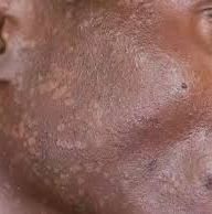
Lichen planus pigmentosus (LPP) is a variant of LP that presents in women over 30 with darker skin. The immunopathogenesis parallels classic LP; however, ultraviolet exposure, cosmetic products, and some medications appear to be key disease modifiers. LPP is also associated with hypothyroidism, hepatitis C, infection, autoimmune disease and malignancy, as well as other LP variants. LPP most commonly affects the neck, followed by face, and then upper extremities, with some sort of facial involvement reported in over half of cases. Symptoms include cosmetic disfigurement, pruritus, and burning, though approximately 30 percent of patients are asymptomatic.
In Fitzpatrick IV-VI, LPP presents as gray-brown macules with irregular, ill-defined borders that gradually coalesce into diffuse, reticulated, perifollicular, or annular hyperpigmented patches. Lesions often involve sun-exposed areas. A subtype, LPP inversus, affects flexural and intertriginous areas not exposed to sunlight. Mechanical friction triggers LPP inversus, and a transition zone from papules to macules is often seen.
When diagnosing LP and LPP, dermoscopy allows for better visualization of the associated dots and globules, as well as any pigmentary changes. The clinician may also see other common associations, such as sparing of follicular openings, telangiectasias, or a blue-white veil. Clinicians should be aware of differentials, including atypical melanocytic lesions, which can be harder to recognize in darker Fitzpatrick skin types. Careful clinicopathologic correlation is required to avoid misdiagnosis. If a biopsy is taken, histopathology will demonstrate a lichenoid inflammatory infiltrate in both LP and LPP. LPP will have a more rapid regression of inflammation, but persistent dermal melanin deposition.
Management is challenging. LPP is frequently refractory to therapy and pigmentary changes may persist for months to years. First-line management includes strict photoprotection. Additional therapies with variable efficacy include topical and systemic corticosteroids, topical tacrolimus, oral vitamin A, azelaic acid, topical retinoids, chemical peels, dapsone, and pigment-targeting lasers. Platelet-rich plasma injections have shown preliminary benefit but require further investigation.


Comments