Molluscum Contagiosum
- UAMS Dermatology Students
- Dec 14, 2025
- 2 min read
Updated: Feb 17
Molluscum contagiosum is a common cutaneous viral infection caused by a DNA poxvirus and is frequently encountered in children and adolescents. Incidence increases during adolescence due to closer skin-to-skin contact, participation in athletics, shared equipment, and shaving practices. The virus infects epidermal keratinocytes, leading to the formation of characteristic dome-shaped papules. Lesions may persist for several months before spontaneous resolution occurs as cell-mediated immunity develops.
Clinically, molluscum contagiosum presents as discrete, smooth, flesh-colored or skin-toned papules with central umbilication. Adolescents may present with numerous papules, often 20–30 lesions distributed across the trunk, extremities, and anogenital regions. Mild pruritus may be present.
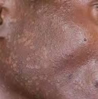
In lighter skin tones, Fitzpatrick I–II, lesions typically appear pink or flesh-colored with a clearly visible central umbilication. Mild inflammatory halos may appear erythematous and are readily appreciated. In Fitzpatrick III–VI, lesions may appear skin-colored, tan, purple, or gray, and dyspigmentation such as hypo- or hyperpigmentation may accompany inflammation or resolution. Dermoscopy can aid diagnosis across typically shows a white or yellow amorphous area with peripheral or branched vessels. Due to inflammation being less visible in darker skin tones, molluscum dermatitis or PIH may be more prominent than the papules themselves. Providers should be aware that that misdiagnosis of molluscum contagiosum commonly occurs as folliculitis, acneiform eruptions, or eczema.
Management strategies depend on lesion burden, symptom severity, cosmetic concerns, and risk of transmission. Because molluscum contagiosum often resolves spontaneously, observation is a reasonable option, although this process may take several months. When treatment is desired, commonly used modalities include cryotherapy or topical agents that promote an inflammatory reaction to enhance immune-mediated clearance. Cryotherapy may cause pigmentation in people with darker skin tones and should be used very judiciously. Extraction and other treatments that protect the melanocytes may be better indicated. In adolescents with darker skin types, minimizing irritation and emphasizing sun protection will help reduce the risk of post-inflammatory pigmentary change, and topical retinoids are one therapeutic option for both treatment and reducing dyspigmentation. It is also of not that destructive therapies such as cryotherapy and cantharidin carry higher risk of dyspigmentation and should be used sparingly. Methods that reduce melanocyte injury, such as curettage and topical retinoids may be preferred in Fitzpatrick IV-VI.

Meza-Romero R, Navarrete-Dechent C, Downey C. Molluscum contagiosum: an update and review of new perspectives in etiology, diagnosis, and treatment. Clin Cosmet Investig Dermatol. 2019;12:373-381. doi:10.2147/CCID.S187224.
Badri T, Gandhi GR. Molluscum contagiosum. In: StatPearls [Internet]. StatPearls Publishing. Updated March 27, 2023. Accessed December 2, 2025. https://www.ncbi.nlm.nih.gov/books/NBK441898/
Hebert AA, Bhatia N, Del Rosso JQ. Molluscum contagiosum: epidemiology, considerations, treatment Options, and therapeutic Gaps. J Clin Aesthet Dermatol. 2023;16(8 Suppl 1):S4–S11.
Dadrass F, Kenner-Bell B. Molluscum contagiosum. Skin of Color Society. Accessed December 2, 2025. https://skinofcolorsociety.org/discover-patientspublic/public-education/molluscum-contagiosum.
Mar K, Khalid B, Maazi M, Ahmed R, Wang OJ, Khosravi-Hafshejani T. Treatment of post-inflammatory hyperpigmentation in skin of colour: a systematic review. J Cutan Med Surg. 2024;28(5):473-480. doi:10.1177/12034754241265716.


Comments