Tinea Versicolor
- UAMS Dermatology Students
- Dec 14, 2025
- 2 min read
Tinea versicolor, also known as pityriasis versicolor, is a superficial fungal infection caused by Malassezia species that commonly affects adolescents due to increased sebum production, physical activity, and humid environmental exposure. Malassezia organisms are part of normal skin flora but convert from their yeast form to their pathogenic hyphal form when exposed to heat, occlusion, and sweat. This transition leads to overgrowth within the stratum corneum and disrupts normal melanocyte activity and melanin distribution, producing the pigmentary variation characteristic of tinea versicolor.
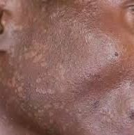
Clinically, tinea versicolor presents as hypo- or hyperpigmented macules and patches with fine scale, most often involving the neck, chest, upper back, and proximal upper extremities. Lesions may be mildly pruritic or asymptomatic, and the condition is frequently recurrent, particularly in adolescents engaged in athletics or living in warm, humid climates.
In lighter skin tones, Fitzpatrick I–III, tinea versicolor often appears as pigmented macules with visible erythema and fine scale. The contrast between affected and unaffected skin is typically pronounced. In Fitzpatrick IV–VI, presentation differs substantially. Lesions may appear as hyperpigmented macules and patches ranging from dark brown to gray-black. The hue may vary among individuals with similar skin types. Erythema may be subtle or absent, and scale can be less apparent. Hypopigmented lesions are often more noticeable on darker skin, prompting earlier evaluation than hyperpigmented variants. Dyspigmentation may persist for weeks to months even after successful antifungal treatment due to delayed normalization of melanocyte activity. Because pigmentary changes are more striking in darker skin, patients may present earlier with concerns. Additionally, providers should be aware that tinea versicolor is misdiagnosed as vitiligo, pityriasis alba, or early cutaneous T-cell lymphoma more frequently in Fitzpatrick IV-VI, partially due to reduced visibility of erythema and scale (chau).
Diagnosis is usually clinical but may be confirmed with Wood’s lamp examination or potassium hydroxide preparation demonstrating the characteristic “spaghetti and meatballs” pattern of hyphae and spores. These tools are particularly helpful in Fitzpatrick IV–VI skin, where pigmentary contrast may be challenging to assess visually.
Treatment includes topical selenium sulfide, zinc pyrithione, or azole-based shampoos and creams applied to affected and surrounding skin. Preventive measures including routine use of antifungal shampoos during warm seasons, minimizing occlusion and sweat retention, and avoiding oily skin products, may help reduce recurrence. In all skin types, especially Fitzpatrick IV–VI, counseling on the slow resolution of pigmentary changes is essential to avoid misinterpretation of persistent dyspigmentation as treatment failure. It is important to educate patients with darker Fitzpatrick skin types that pigmentary abnormalities frequently outlast the fungal infection. Additionally, the importance of sun protection should be noticed as to reduce contrast while melanocyte function normalizes.


Comments